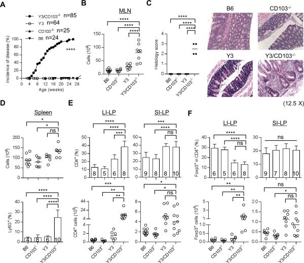
Figure 1. Y3/CD103−/− mice develop severe colitis.
(A) Incidence of disease. Mice from each genotype were monitored weekly for signs of colitis, and were considered diseased when they exhibit at least one of the following severe diarrhea, rectal prolapse and/or wasting. (B) MLN cellularity. (C) Histology scoring of the large intestine of age-matched 12-24 week-old mice (left). Formalin-fixed and H&E stained colon sections (right) were examined. Typically, 1 = mild inflammatory infiltrates, 2 = moderate, 3 = extensive, with or without necrosis (n ≥ 3 mice/group), (original magnification 12.5 X). (D) Splenic cellularity (top) and the frequency of Ly6G+ granulocytes (bottom) in the spleen were assessed by flow cytometry. (E, F) The proportion and number of total CD4+ (E) and CD4+ Foxp3+ Tregs (F) in the LI-LP and SI-LP were determined by flow cytometry. (B, D-F) Age-matched 12-24 week-old mice were assessed for each genotype. The number of mice/group is indicated within each bar or by individual symbols in scatter graphs.

