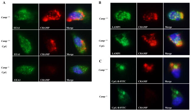
Figure 8. CRAMP colocalizes with CpG in the endolysosome and associates with TLR9.
A, WT and Camp−/− Flt3L cDCs were stimulated with or without 1 μM CpG-B for 90 min. Cells were fixed, permeabilized and stained for CRAMP (Alexa 568; red) and EEA1 (Alexa 488; green, A). Nuclei were stained with DAPI (blue). The localization of CRAMP was analyzed by fluorescence microscopy. B, WT Flt3L cDCs were stimulated with or without 1 μM CpG-B for 90 min. Cells were fixed, permeabilized and stained for CRAMP (Alexa 568; red) and LAMP1 (Alexa 488; green, B). C, WT and Camp−/− Flt3L cDCs were stimulated with 1 μM FITC-labelled CpG-B for 90 min. Cells were fixed and stained for CRAMP (Alexa 568; red).

