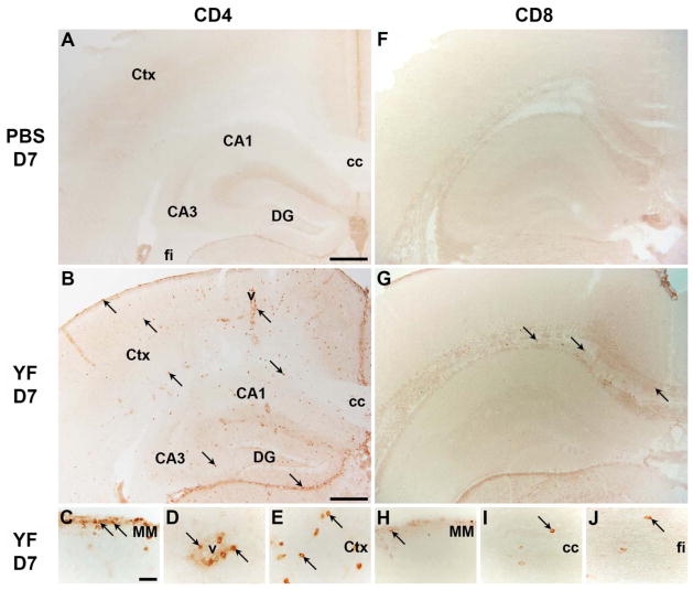
Figure 5. Immunohistochemical analysis of CD4+ and CD8+ T cells into YF-17D infected brains.
A–E) Immunohistochemical analysis of CD4 showed absence of CD4+ cells in brains of PBS injected mice (A), while CD4+ cells had migrated into the brains of YF-17D i.c. infected mice day 7 post injection (B–E). F–J) Similarly, immunohistochemical analysis of CD8 showed absence of CD8+ cells in brains of PBS injected mice (F), while CD8+ cells had migrated into the brains of YF-17D i.c. infected mice day 7 post injection (G–J). vCA1-3: cornu ammonis 1 and 3; cc: corpus callosum; Ctx: cortex; DG: dentate gyrus; v: vessel; MM: meningeal membrane; fi: fimbria. Scale bar: (A, B, F, G) 400 μm, (C–E, H–J) 50 μm. Sections stained with control abs are depicted in a supplementary figure (Suppl. Fig. S1).

