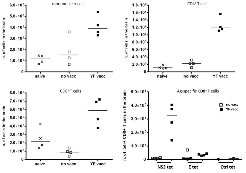
Figure 8. Composition of mononuclear cells isolated from YF-17D infected brains.
WT B6 mice that were either naïve or had been immunized s.c. with 105 pfu of YF-17D virus 28 days earlier were challenged with 104 pfu of YF-17D virus i.c., and 5 days later cells were isolated from the brains; naïve mice served as controls. Absolute numbers of mononuclear cells, CD4+, CD8+ and YF specific CD8+ cells are depicted. Antigen-specific CD8+ T cells were detected by tetramer staining using NS3(268-275) or E(4-12) PE and APC-conjugated tetramers (tet); LCMV specific PE and APC-conjugated tetramers were used as controls (Ctrl tet). Each dot represents pooled cells from 2 mice.

