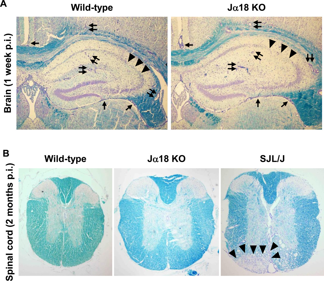
Figure 1.
Neuropathology of wild-type C57BL/6 and Jα18 knockout (KO) mice, 1 week and 2 months post infection (p.i.). Wild-type C57BL/6 and Jα18 KO mice were infected intracerebrally with the Daniels (DA) strain of Theiler’s murine encephalomyelitis virus (TMEV). (A) During the acute phase of TMEV infection, 1 week p.i., wild-type C57BL/6 and Jα18 KO mice had similar levels of meningitis (arrows), perivascular cuffing (paired arrows), and neuronal loss (arrowheads) in the hippocampus. Sections were representatives of six wild-type C57BL/6 and 10 Jα18 KO mice. Magnification, 35×. (B) During the chronic phase of TMEV infection, 2 months p.i., neither wild-type C57BL/6 nor Jα18 KO mice developed lesions in the spinal cord, while susceptible SJL/J mice developed severe demyelination (arrowheads) in the ventral funiculus of the spinal cord. Sections were representatives of 12 wild-type C57BL/6 and 21 Jα18 KO mice. Magnification, 23×. (A, B) Luxol fast blue staining.

