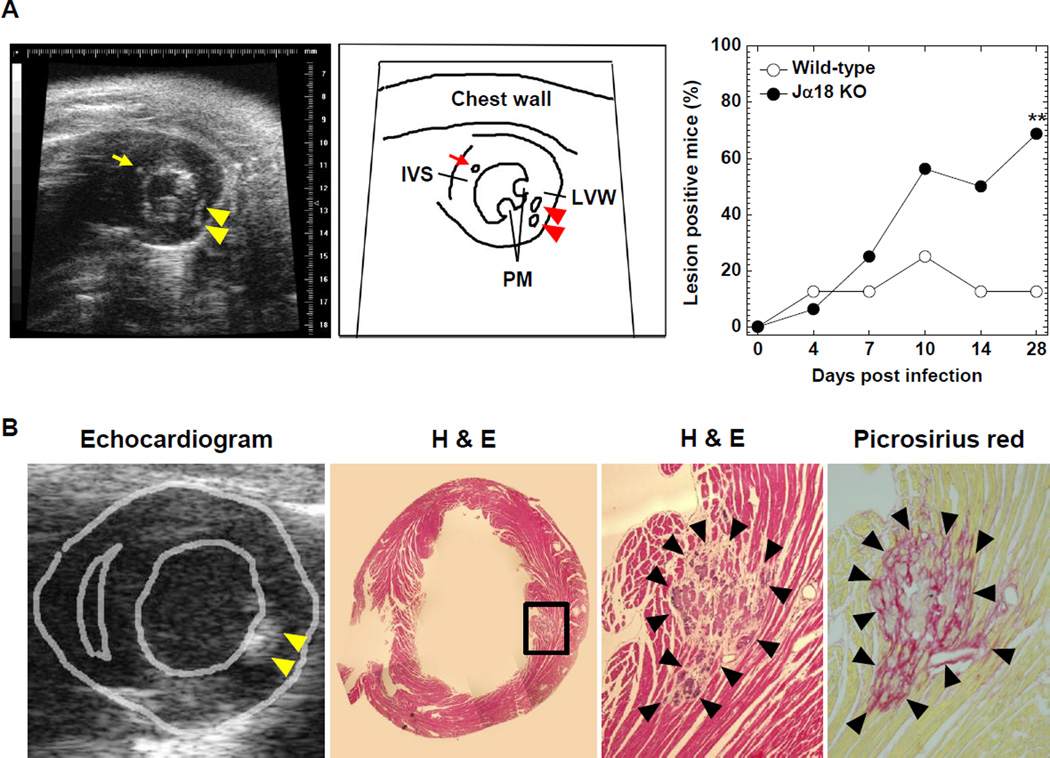
Figure 6.
Echocardiograms and heart sections stained with H & E or picrosirius red of mice infected with TMEV. (A) The left and middle panels show the short axis view at the papillary muscle (PM) level in an echocardiogram and the scheme, respectively. Some TMEV-infected wild-type C57BL/6 and Jα18 KO mice had high intensity lesions in the interventricular septum (IVS, arrow) and left ventricular wall (LVW, arrowheads). The incidence of mice with high intensity lesions in the heart was higher in Jα18 KO mice, compared with wild-type C57BL/6 mice during the 1-month observation period (**P < 0.01, χ2 test). (B) H & E and picrosirius red staining (magnification, 52×) showed that high intensity lesions in echocardiograms corresponded to basophilic degeneration, calcification, and fibrosis in the hearts of mice with TMEV-induced myocarditis. The picture showing the morphology of the heart (second from the left) is a composite picture. (A, B) Echocardiograms are representative of 8 to 16 mice/group/time point.

