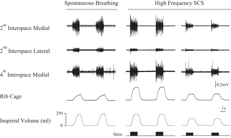
Fig. 1.
Multiunit EMG recordings form the parasternal intercostal muscles during spontaneous breathing and high-frequency spinal cord stimulation (HF-SCS) in one animal. From top to bottom, tracings represent multiunit EMG recordings of the parasternal intercostal muscles from the medial bundles of the 2nd interspace, lateral bundles of the 2nd interspace, and medial bundles of the 4th interspace and inspired volume. Recordings were obtained during spontaneous breathing (left panel) and HF-SCS with matching of inspired volume to spontaneous breathing (middle panel) and during HF-SCS with matching of rib cage expansion to spontaneous breathing (right panel). See text for further explanation.

