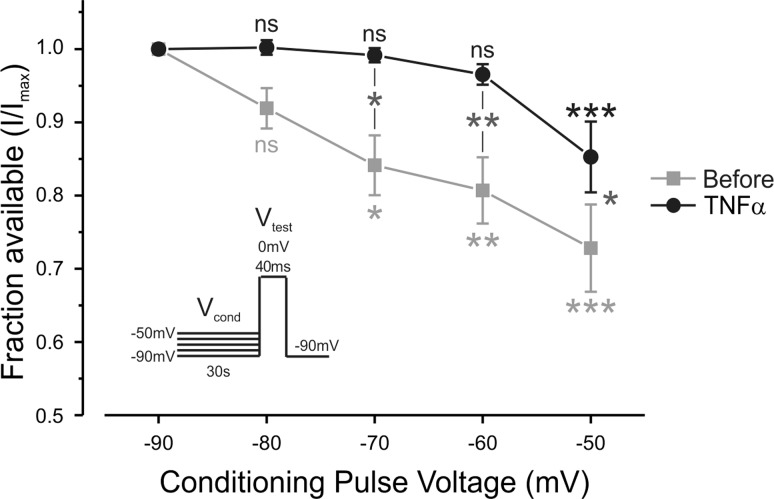
Fig. 11.
TNF-α relieves TTX-r channels from ultraslow inactivation at hyperpolarized potentials. A fraction of channels available for activation (peaks of TTX-r sodium currents during a step to 0 mV, normalized to Imax, means ± SE) following prolonged 30-s holding at voltages, as indicated in the x-axis, before (gray squares) and 3 min after application of TNF-α (black circles). Currents were elicited by 10-ms test steps to 0 mV, after 30-s conditioning pulses (Vcond) to the indicated voltages. Note that the substantial current reduction, following prolonged holding at Vcond of −70 mV, −60 mV and −50 mV (black asterisks, comparison within Before group, ns, P > 0.05, **P < 0.01, ***P < 0.001 one-way ANOVA with post hoc Bonferroni, n = 6), was diminished by application of TNF-α (gray asterisks, comparison within TNF-α group, ns, P > 0.05, ***P < 0.001, one-way ANOVA with post hoc Bonferroni, n = 6). Note also that TNF-α leads to increase of available channels at −70 mV by about 15%. Light great asterisks, comparison between Before and TNF-α groups. *P < 0.05; **P < 0.01, two-way ANOVA with post hoc Bonferroni, n = 6. Inset: stimulation protocol.

