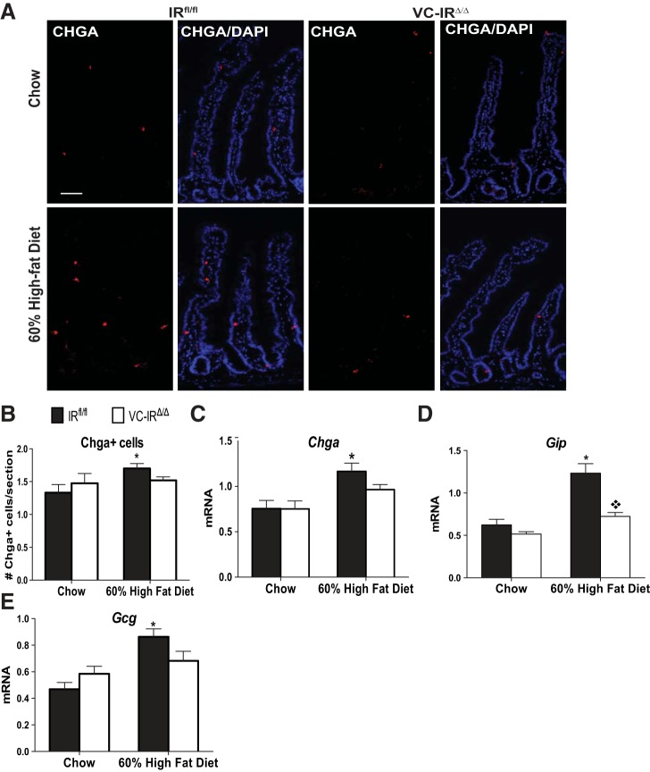
Fig. 7.
IEC-IR loss prevents HFD-induced increases in chromogranin A (CHGA)-positive cells and Chga, glucose-dependent insulinotrophic peptide (Gip), and glucagon (Gcg) mRNAs. A: representative images of CHGA immunostaining (red) and DAPI (blue). Magnification ×20; scale bar = 50 μm. B: quantification of number of CHGA-positive enteroendocrine cells in chow- or HFD-fed IRfl/fl and VC-IRΔ/Δ mice from images in A. C–E: quantitative RT-PCR assessment of Chga (C), Gip (D), and Gcg (E) mRNA levels in jejunal epithelial cells isolated from chow- or HFD-fed IRfl/fl and VC-IRΔ/Δ mice. Data are normalized to β-actin invariant control. Values are means ± SE; n ≥ 6. *P < 0.05 (by 2-way ANOVA with Sidak's post hoc pair-wise comparisons for main effect of diet). ◇P < 0.05 (by 2-way ANOVA with Sidak's post hoc pair-wise comparisons for main effect of genotype).

