Fig. 9.
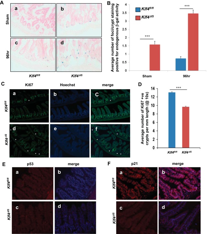
A: staining for senescence-associated endogenous β-galactosidase (β-gal) activity in the intestines of Klf4fl/fl (a and c) and Klf4ΔIS (b and d) mice. B: quantification of β-gal foci per crypt in Klf4fl/fl and Klf4ΔIS mice at sham and 96 h following γ-irradiation. The number of positive β-gal foci per crypt was averaged from at least 20 crypts per mouse. C: analysis of mice intestines at 7–9 days postirradiation of Klf4fl/fl (a–c) and Klf4ΔIS (d–f) mice. Atrophied crypts are marked by asterisks. D: quantification of the percentage of atrophied crypts per field in Klf4fl/fl and Klf4ΔIS mice at 7–9 days following γ-irradiation. The number of atrophied crypts was averaged from at least 10 fields per mouse. E: p53 staining of Klf4fl/fl (a and b) and Klf4ΔIS (c and d) mice. p53 is red, and nuclear staining is blue. F: p21 staining of Klf4fl/fl (a and b) and Klf4ΔIS (c and d) mice. p21 is red, and nuclear staining is blue; n = 4, ***P < 0.001 ± SE.

