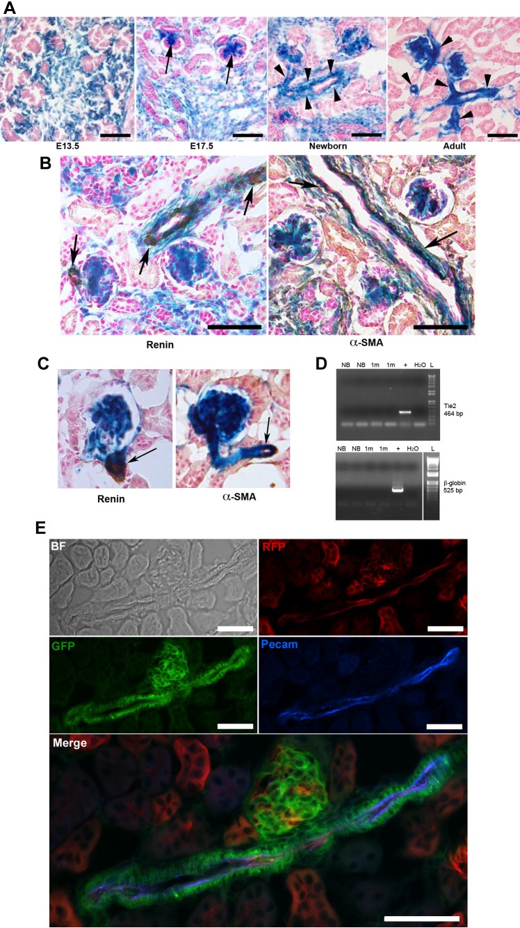
Fig. 2.
Foxd1+ cells are an early precursor for mesangial, vascular smooth muscle, renin cells, and pericytes but not for endothelial cells. A: Foxd1cre;R26R kidneys sections at different developmental stages showing extensive expression of β-gal within the undifferentiated stromal compartment (E13.5), with progressive differentiation into glomerular mesangium (E17.5, arrows), and cells within the wall of the arterial tree (newborn and adult, arrowheads). Endothelial cells within the arterioles do not express β-gal. B: Foxd1cre;R26R newborn kidneys sections showing coexpression of β-gal with immunostaining for renin (left, brown) and with α-SMA (right, brown). Endothelial cells do not express β-gal. C: immunostaining for renin (left) and α-SMA (right) in kidney sections from adult mice show coincidence of the DAB product (brown) with the X-gal staining (blue) (arrows). D: RT-PCR for Tie2 (top) and β-globin (bottom) was positive in samples of whole kidneys (+) and negative in FACS isolated Foxd1-lineage cells from Foxd1cre;mTmG newborn (NB) and 1-mo-old (1m) mouse kidneys. L, 1KB ladder; H2O, negative control. E: immunofluorescence for Pecam in kidney section from an adult Foxd1cre;mTmG mouse shows lack of coincidence of the blue fluorescent marker with cells derived from the Foxd1 precursors (GFP). However, ECs identified by Pecam expression are also positive for RFP, which marks cells not derived from Foxd1 progenitors. Scale bars: 50 μm.

