Fig. 5.
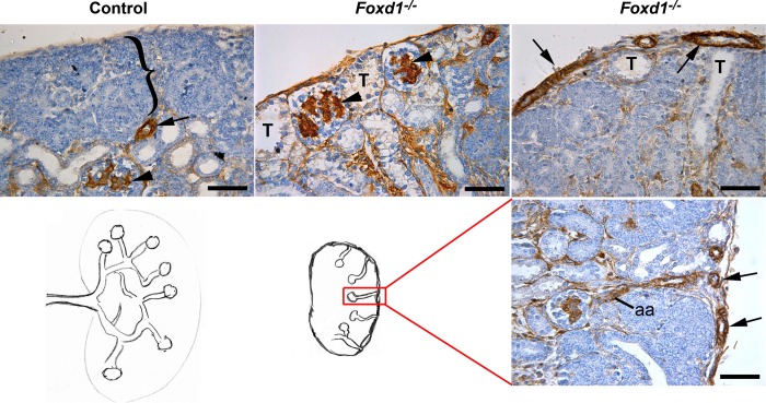
Foxd1−/− kidneys display abnormal topological orientation of the renal vessels and disorganized renal structure. Immunostaining for α-SMA (brown) of E17.5 kidneys show that control mice present a normal outer undifferentiated nephrogenic zone (bracket) and a further developed inner cortex with arterioles (arrow) and more mature glomeruli expressing α-SMA in the mesangium (arrowhead), whereas Foxd1−/− mice present abnormal subcapsular glomeruli (arrowheads) and tubules (T) and capsular arteries (arrows). Lower panel shows a drawing of the vascular pattern in control and Foxd1−/− mice, as seen in the section stained with α-SMA antibody to highlight the abnormal arteries (arrows) and afferent arterioles (aa). Scale bar: 50 μm.

