
Figure 1.
Shox2 Is Specific to the Developing Mouse SAN
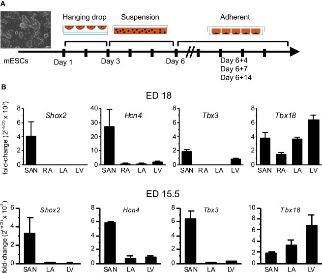
(A) A schematic diagram of cardiac differentiation protocol. mESCs were dissociated into single cells on day 1(D1) and cultured in suspension for 2 days in hanging drops and then 3 days on a nonadhering culture plate. On D6, the cells were plated on an adherent plate. The differentiating cells were characterized 4, 7, and 14 days from D6.
(B) Transcript expression levels of the transcription factors that govern cardiac pacemaker cell specification were quantified by real-time RT-PCR from microsurgically isolated SAN tissues from ED 18 mouse hearts (top). The bottom illustrates the gene expression level at ED 15.5. Expression analysis of RA was not performed at ED 15.5 because the tissue was too small to be reliably separated from the SAN. Purity of the SAN preparation is validated by robust Hcn4 expression limited to the SAN (second from left). Data are represented as mean ± SEM. All experiments were performed in three independent biological replicates.