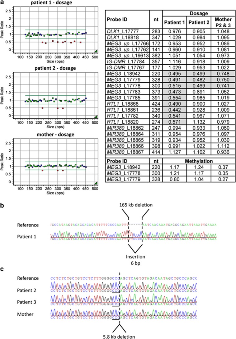
Figure 3.
Results of the (a) MS-MLPA analysis and (b, c) the Sanger sequencing of the junction fragments obtained by long-range PCR for breakpoint identification. (a) The MS-MLPA for patient 1 showed a reduced dosage of about 50% for the nine probes encompassing MEG3, RTL1 and part of the miRNA cluster confirming the presence of a heterozygous deletion (plot and table). The probes for DLK1 and (the two probes) next to the IG-DMR displayed a normal dosage (around 1.0). A strong hypermethylation was demonstrated by the methylation-specific probes for the MEG3-DMR indicating that the deletion occurred on the unmethylated maternal allele (lower part of the table; normal methylation value should be ∼0.5). Results for the patients' mother were normal (not shown), so that the deletion seemed to have occurred de novo. For patients 2 and 3 the dosage of three MLPA probes for the 5′ end of MEG3 are reduced by ∼50% indicating the presence of a heterozygous deletion (plot and table). Dosage reduction in the mother was ∼30%, pointing to a mosaic state of the deletion. Results of the methylation-specific probes showed a strong hypermethylation in the two patients and a slight hypomethylation of about 36% in the mother, who therefore carries the deletion on the paternal allele (Data for patient 3 not shown). (b) and (c) Sanger sequencing results for the deletion breakpoints. For patient 1 a 165 kb deletion comprising a 6 bp insertion between the proximal and distal breakpoints was observed (chr14:g.101285913_101451066del, hg19). For patients 2 and 3 as well as their mother a 5.8 kb deletion was present. The breakpoint junctions are uncertain at the triple C position (underlined; chr14:g.101291322_101297145del, hg19).

