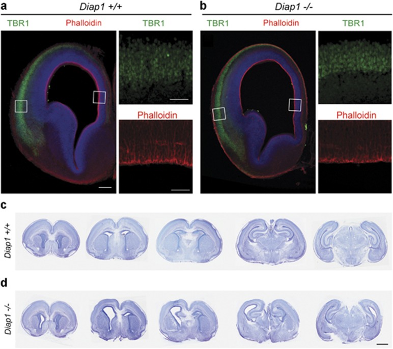
Figure 4.
Cortical organization is preserved in the Diap1−/− mouse. Image depicts deeper cortical layer marker TBR1, layer VI (green), and phalloidin staining in the coronal sections of the lateral ventricle wall in (a) Wt (Diap1+/+) and (b) mutant (Diap1−/−) mice at E14.5 day. Boxed areas are shown at the higher magnification. Scale bars 200 μm at lower magnification and 50 μm at higher magnification. (c–d) Nissl staining of coronal brain sections of Diap1-KO and Wt at P0. Note the lateral ventricle dilatation on the Diap1-KO mouse. Scale bar is 1 mm.

