
Figure 3. Mdivi-1 enhances death receptor-mediated apoptotic signaling.
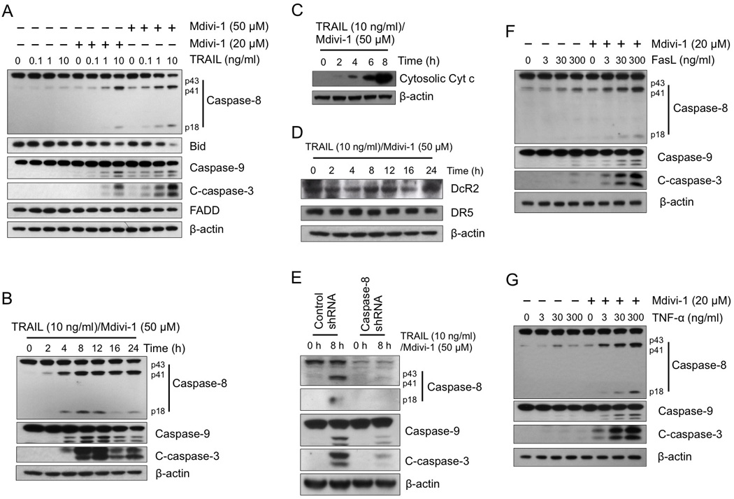
(A) A2780 cells were treated with TRAIL alone, mdivi-1 alone, or the combination at the indicated concentrations for 16 h. The activation of apoptotic signaling was examined by western blot using antibodies against caspase-8, bid, caspase-9, cleaved caspase-3 (C-caspase-3), and FADD. β-actin was used as a loading control. (B) A2780 cells were treated with the combination of TRAIL and mdivi-1 as indicated. Cleavage of caspases was detected by western blot. (C) A2780 cells were treated with the combination of TRAIL and mdivi-1 as indicated with the presence of 20 µM caspase inhibitor Q-VD-OPH. The cytosolic fraction was isolated using digitonin permeabilization followed by centrifugation. The amount of cytochrome c presented in cytosolic fraction was detected by western blot. (D) A2780 cells were treated as indicated and the expression of Decoy receptor DcR2 and death receptor DR5 were detected by western blot. (E) A2780 cells were transduced with control or caspase-8 shRNA and selected by puromycin. Cells were treated with the combination of TRAIL and mdivi-1 at indicated concentration for 8 h. The cleavage of caspases was detected by western blot. (F, G) A2780 cells were treated with increasing doses of Fas ligand (FasL) (F) or TNF-α (G) alone or the combination with mdivi-1 (20 µM) for 16 h. Cleavage of caspases was examined by western blot. These data represent three independent experiments.