
Fig 5.
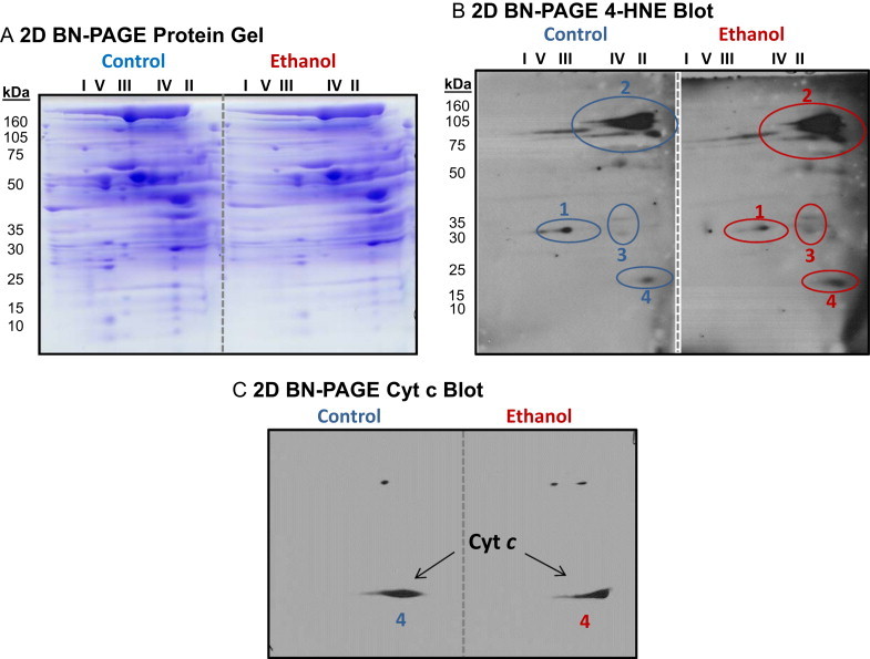
High-resolution separation of liver mitochondrial proteins from control and ethanol fed rats using 2D BN-PAGE and 4-HNE detection by western blot. Panel A: Representative 2D BN-PAGE gels of liver mitochondrial proteins from one pair of control and ethanol-fed rats. BN-PAGE was performed as described in methods sections. Panel B: Corresponding 2D 4-HNE western blots are shown for gels in panel A. Four protein spot clusters showed immunoreactivity for 4-HNE in mitochondria isolated from livers of control (C) and ethanol (E) fed rats. Three of the four protein spots (1–3) were identified by mass spectrometry (Table 2). Note that for protein spot cluster no. 3 the bottom spot is enoyl CoA hydratase and the top spot is most likely β-hydroxybutyrate dehydrogenase; however, MOWSE score was too low for positive ID. panel C: Immunoblot for cytochrome c from control and ethanol fed mitochondrial samples separated using 2D BN-PAGE. The protein spot immunoreactive for cytochrome c co-localizes to protein spot 4 in panel B. Previous studies from our laboratory show no difference in the amount of total cytochrome c protein in liver mitochondria isolated from control and ethanol-fed rats [8].