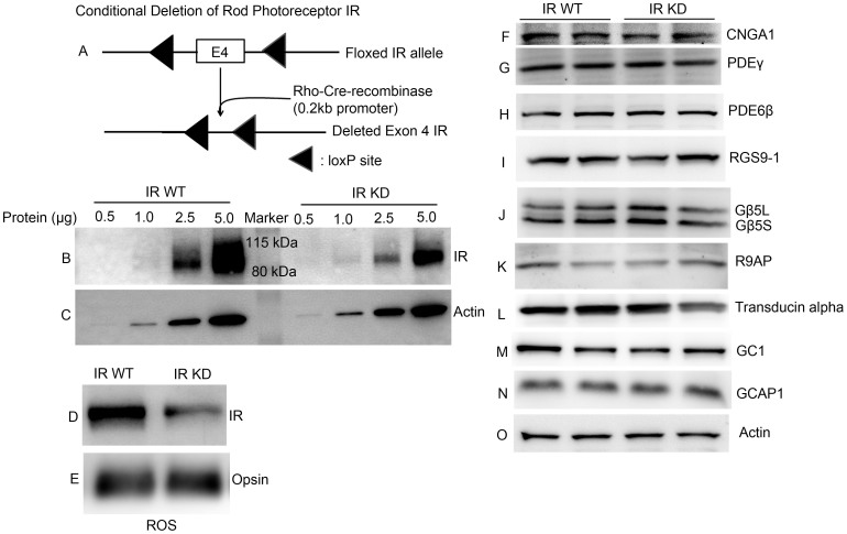
Figure 1. Expression levels of transduction proteins in wild type and IR knock-down animals.
(A) Schematic diagram of loxP floxed IR loci. Rod-photoreceptor-specific IR knock-down mice were generated by breeding mice with a floxed IR with mice that express Cre recombinase under the control of rod opsin promoter (0.2 kb). To determine the deletion of IR, varying amounts of retinal proteins (0.5, 1.0, 2.5 and 5.0 μg) from IR wild type and IR knock-down (IR-KD) mouse retinas were subjected to immunoblot analysis with antibodies against (B) IR and (C) actin. Rod outer segments (ROS) were prepared by discontinuous sucrose (47%, 37% and 32%) density gradient centrifugation (see Methods). ROS from IR wild type and IR knock-down mice were immunoblotted with (D) anti-IR and (E) anti-opsin antibodies. Ten micrograms of two independent retinal proteins were subjected to immunoblot analysis with antibodies against (F) CNGA1, (G) PDEγ, (H) PDE6β, (I) RGS9-1, (J) Gβ5L (also detects short form Gβ5S), (K) R9AP, (L) transducin alpha, (M) GC1, (N) GCAP1, and (O) actin. For Fig. 1F, the blot is a reprobe of the gel used for PDE6 β (H); that is, after the PDE6 β measurement, the gel was stripped and reprobed with an anti-CNGA1 antibody. Full-length blots/gels are presented in Supplementary Figure 1.

