FIGURE 1.
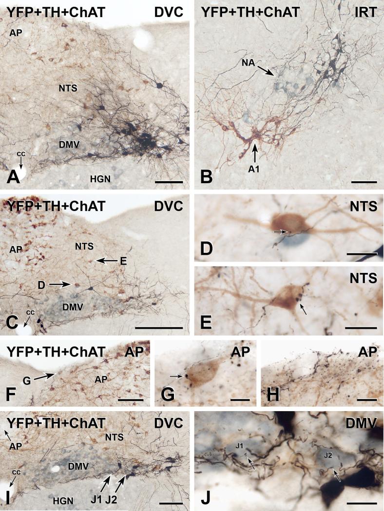
Three-colour immunoperoxidase labelling for YFP in YFP-PPG neurons (black), tyrosine hydroxylase (TH) in catecholamine neurons (brown) and choline acetyltransferase (ChAT) in cholinergic neurons (blue-grey) in transverse sections through the dorsal medulla of YFP-PPG mice. A, Low magnification micrograph showing the cell bodies of black, YFP-immunoreactive PPG neurons in the nucleus of the solitary tract (NTS). These neurons lie lateral to the dorsal motor nucleus of the vagus (DMV) and the majority occur at the level of the area postrema (AP). The dendrites of the YFP-PPG neurons in NTS extend dorsomedially and also run lateral to the hypoglossal nucleus (HGN) and along the border between the DMV and the HGN. cc, central canal. Bar, 100 μm. B, In the intermediate reticular nucleus (IRT), the black, YFP-immunoreactive cell bodies of PPG neurons are located dorsomedial to both the brown, TH-immunoreactive neurons of the A1 cell group and the blue-grey, ChAT-immunoreactive neurons of the nucleus ambiguus (NA). Bar, 100 μm. C, Low magnification micrograph showing the dorsal vagal complex at the level of the most rostral YFP-PPG neurons. A collection of YFP-immunoreactive dendrites travels between the DMV and HGN. Arrows, brown, TH-immunoreactive cell bodies that are shown at higher magnification in D and E. Bar, 250 μm. D and E, Single varicosities (arrows) of black, YFP-immunoreactive axons closely appose brown, TH-immunoreactive cell bodies in the NTS. Bars, 20 μm. F, Low magnification micrograph showing the AP near the level of the most rostral YFP-PPG neurons. Arrow, a brown, TH-immunoreactive cell body near the surface of AP that is shown at higher magnification in G. Bar, 100 μm. G, A single varicosity (arrow) of a black, YFP-immunoreactive axon closely apposes the brown, TH-immunoreactive cell body in AP. Bar, 10 μm. H, At the surface of rostral AP, black, varicose, YFP-immunoreactive axons and black, varicose, YFP-immunoreactive dendrites are intermixed with brown, TH-immunoreactive dendrites arising from AP neurons. Bar, 20 μm. I, Low magnification micrograph showing the DMV near the level of the most rostral YFPPPG neurons. Arrows J1 and J2, two blue-grey, ChAT-immunoreactive vagal motor neurons that are shown at higher magnification in J. Bar, 100 μm. J, Fine varicosities (arrows) of black, YFP-immunoreactive axons closely appose the blue-grey, ChAT-immunoreactive cell bodies of DMV neurons J1 and J2. Bar, 20 μm.

