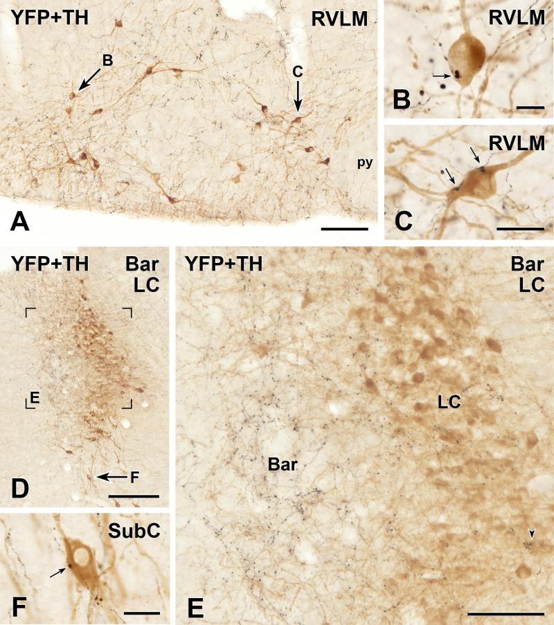
FIGURE 3.
Two-colour immunoperoxidase labelling for YFP in YFP-PPG neurons (black) and tyrosine hydroxylase (TH) in catecholamine neurons (brown) in transverse sections through the rostral ventrolateral medulla (RVLM) and dorsal pons of YFP-PPG mice. A, Low magnification micrograph showing the RVLM. Arrows, brown, TH-immunoreactive cell bodies that are shown at higher magnification in D and E. py, pyramidal tract. Bar, 100 μm. B and C, Single varicosities (arrows) of black, YFP-immunoreactive axons closely appose brown, TH-immunoreactive cell bodies in the RVLM. Bar in B, 10 μm; bar in C, 20 μm. D, Low magnification micrograph showing the region of the locus coeruleus (LC) and Barrington’s nucleus (Bar). Left is medial and the ventricle appears at top left. The region in Box E is shown at higher magnification in E. Arrow F, a brown, TH-immunoreactive neuron in the subcoeruleus region (SubC) that is shown at higher magnification in F. Bar, 250 μm. E, There are many black, varicose YFP-immunoreactive axons in Bar. However, only a few, black, varicose YFP-immunoreactive axons travel amongst the brown, TH-immunoreactive neurons of LC. Of all of the TH-immunoreactive cell bodies in this region of LC, only 6 received close appositions from YFP-immunoreactive varicosities, one of which (lower right) is marked by an arrowhead. There were no close appositions on the TH-immunoreactive dendrites of LC neurons that penetrated Bar. Bar, 50 μm. F, A single varicosity (arrow) of a black, YFP-immunoreactive axon closely apposes the brown, TH-immunoreactive cell body in SubC. Bar, 20 μm.

