
Figure 1.
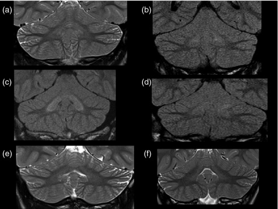
Patients with EAST syndrome show increased signal intensity of the cerebellar nuclei and cerebellar hypoplasia on brain magnetic resonance imaging. Coronal T2 or fluid-attenuated inversion recovery images show varying degrees of signal hyperintensity in the cerebellar deep nuclei. These ranged from subtle signal changes in comparison with the cerebellar cortex to more obvious changes within both the cerebellar deep nuclei and hilar white matter. (a) patient 1-1, (b) patient 2, (c) patient 4, (d) patient 3, (e) patient 5-1, and (f) patient 5-2.