
Figure 2.
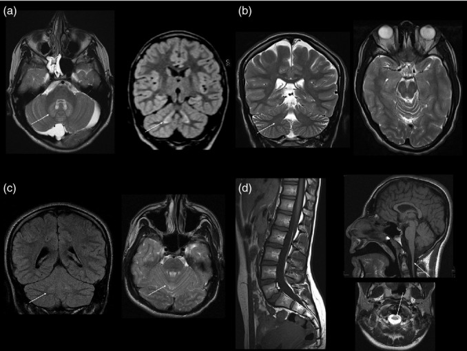
Other abnormalities on brain magnetic resonance imaging. (a) Axial T2-weighted and coronal volumetric fluid-attenuated inversion recovery images in patient 3 show bilateral symmetrical signal hyperintensity within the cerebellar deep nuclei and hilar white matter (arrow) and a retrocerebellar arachnoid cyst. This child also has a fatty filum terminale but normal position of the conus medullaris and no other spinal dysraphic features (not shown). (b) Coronal and axial T2-weighted images in patient 5-2 show prominence of the cerebellar hemisphere and vermian fissures. Note again the subtle symmetrical dentate nucleus changes (arrow). (c) Sagittal T1-weighted image (patient 1-1) shows normal position of the conus medullaris, a fatty filum terminale, and normal spinal cord volume. (d) Sagittal T1- and axial T2-weighted brain images (patient 2) show global lack of cerebral volume with slightly thin corpus callosum and brainstem and spinal cord hypoplasia.