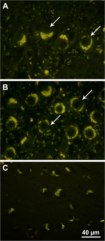
Figure 2.

Fluorescence micrographs of brain sections of cerebellum (A and B) and cerebral cortex (C) demonstrating the massive intracellular accumulation of yellow-emitting autofluorescent storage bodies. In the cerebellum the storage body accumulation was most pronounced in the Purkinje cells (arrows in A and B). The section in (A) was cut perpendicular to the plane of the Purkinje cell layer and the section in (B) was cut parallel to the plane of the Purkinje cell layer. The storage body accumulation in the neurons of the cerebral cortex was perinuclear and asymmetric. Bar in (C) indicates the magnification for all 3 micrographs.
