
Figure 4.
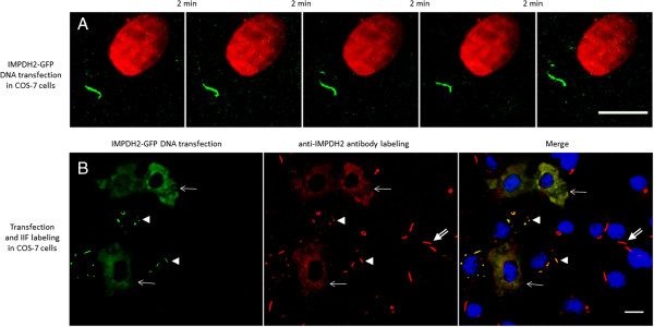
IMPDH2-GFP-tagged RR induced in live COS-7 cells are detected in stable and stationary state. COS-7 cells were transfected with IMPDH2-GFP plasmid and maintained in medium containing 1 mM ribavirin for 18 h. (A) Sequential pictures were captured for >10 min and representative images are shown at 2 min intervals. Similar to the antibody labeling experiment, changes in shape and size of RR were not observed. (B) Transfected cells were fixed and labeled by IIF with rabbit anti-IMPDH2 antibody (red). Cells with a high level of transfection (arrows, left panel) do not show RR (arrows). Cells with low transfection levels show RR (arrowheads). Of 336 cells counted, 16% show IMPDH2-GFP RR; transfection efficiency was 37%. Cells not transfected by IMPDH2-GFP showed RR labeled by anti-IMPDH2 antibody (double-arrows). All transfected IMPDH2-GFP-tagged RR (green, arrowheads) were labeled by rabbit anti-IMPDH2 antibody (red). Nuclei were counterstained with DAPI (blue). Data represent three in (A) and two in (B) independent experiments. Bars: 10 μm.