
Figure 1.
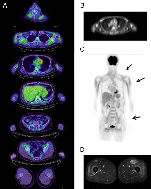
[18F] fluorodeoxyglucose positron emission tomography (FDG PET) and MRI findings in a patient with dermatomyositis (patient 11). Serial sections of FDG PET/CT (A). Using FDG uptake in mediastinum blood vessels as a positivity criterion, positive regions were found in the paraspinal muscles, shoulders, upper arms and lumbar girdles in a predominantly symmetrical distribution. An FDG PET image of the upper arm level (B). Frontal view of an FDG PET image indicating FDG uptake in the shoulders, upper arms and iliopsoas (C). MRI of the thighs (D) showing high signal areas in the bilateral quadriceps femoris (T2-weighted images with fat suppression).