
Figure 3.
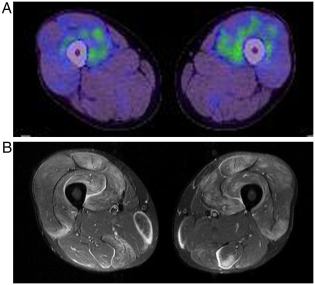
[18F] fluorodeoxyglucose positron emission tomography (FDG PET) (A) and MRI findings (B) for both thighs in a patient with polymyositis (patient 14). Distribution patterns of high signal on MRI and FDG PET are different. The FDG uptake is localised and predominantly within the muscles.