Figure 2.

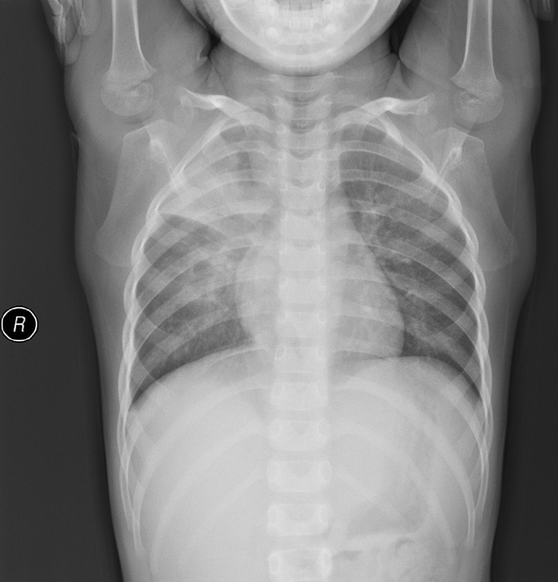
An example of right upper and middle lobe infiltration from a chest radiograph obtained 5 days after the onset of symptoms in a patient with mycoplasma pneumonia.

An example of right upper and middle lobe infiltration from a chest radiograph obtained 5 days after the onset of symptoms in a patient with mycoplasma pneumonia.