Figure 4.
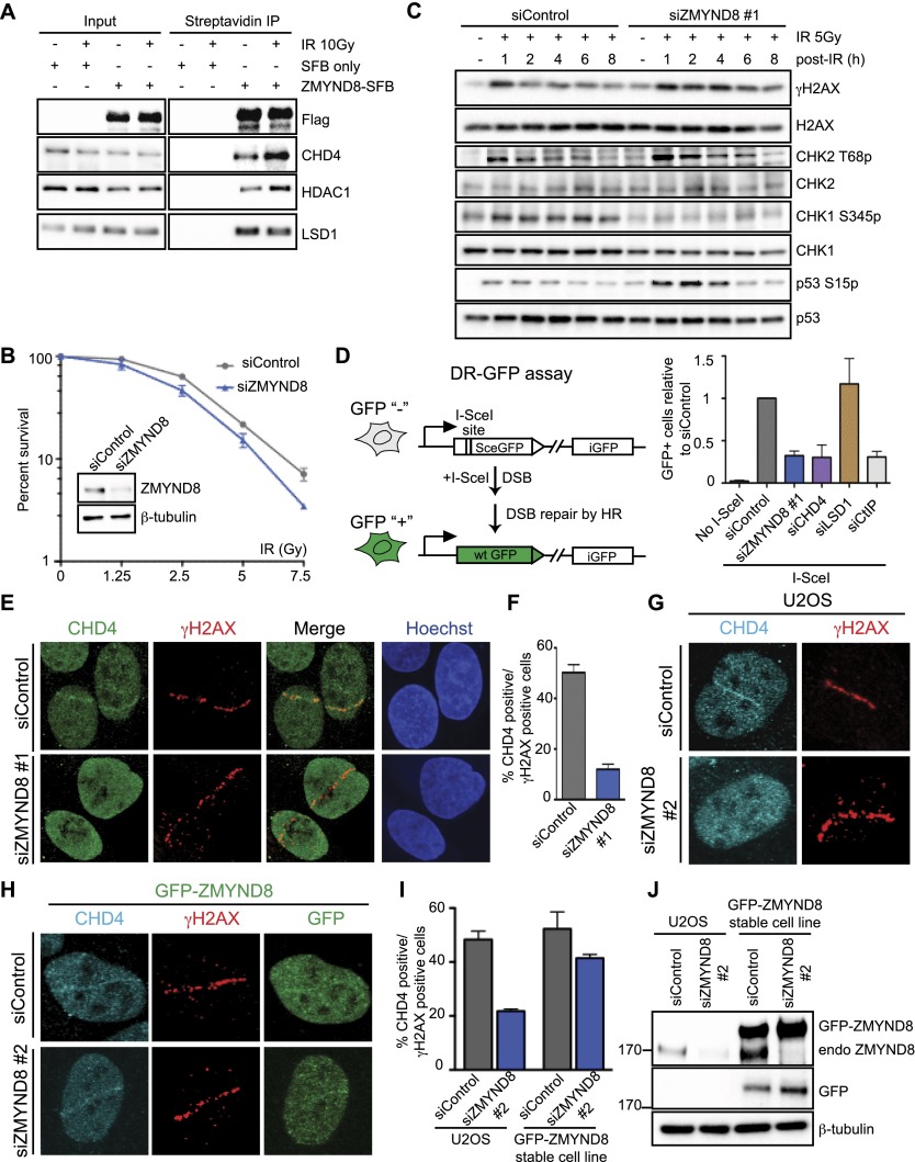
ZMYND8 participates in the DDR and recruits CHD4 to damaged chromatin. (A) ZMYND8 interacts with CHD4 upon IR treatment. Experiments were performed as in Figure 3E with IR. (B) Clonogenic assays reveal hypersensitivity of ZMYND8-depleted cells to IR. U2OS cells were treated with control or ZMYND8 siRNAs and damaged with various doses of IR. Graphs are mean ± SEM; n = 2. (C) ZMYND8-depleted cells are defective in DNA damage signaling. siControl and siZMYND8 U2OS cells were IR-treated and analyzed at the indicated time points by Western blotting. Several phosphorylated DNA damage markers were analyzed with unmodified antibodies acting as loading controls. (D) Cells depleted of ZMYND8 and CHD4, but not LSD1, are defective in HR. DR-GFP reporter assays were performed after depletion of the indicated proteins by siRNAs. Depletion of CtIP was used as a positive control. Error bars indicate SEM; n = 3. (E–G) Recruitment of CHD4 to laser damage requires ZMYND8. siControl and siZMYND8 U2OS cells were laser-damaged, and endogenous CHD4 accumulation was analyzed by immunofluorescence. (F) Quantification of E. Data were obtained from >50 cells from three independent experiments. Error bars indicate mean ± SEM. (G) The same results obtained as in E using an independent siRNA targeting the 3′ untranslated region (UTR) of ZMYND8. (H–J) Ectopically expressed GFP-ZMYND8 rescues defective CHD4 damage accrual in ZMYND8-depleted cells. U2OS cells stably expressing GFP-ZMYND8 were treated with si3′ UTR targeting endogenous but not GFP-tagged ZMYND8, which lacks the 3′ UTR. (I) Quantification of G and H performed as in F. n = 2. (J) Western blot analysis of samples from G and H with the indicated antibodies. Note: 3′ UTR siRNA targeting ZMYND8 depletes endogenous but not GFP-tagged ZMYND8.

