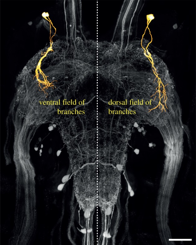
Figure 3.

Partial reconstruction of a pair of neurons (T3-3 and T3-4) in the metathoracic ganglion that show increased serotonin expression following exposure to all gregarizing stimuli. The neurons have a ventral field of branches in the VAC and a dorsal field of branches projecting along the dorso-lateral edge of the neuropile. The reconstructions (yellow) are superimposed on a projection view of the serotonin immunofluorescence (grey) in the entire ganglion. In the reconstructions, the dorsal field of arborizations has been omitted in the left half of the ganglion (to show only the somata, primary neurites and ventral field), and the ventral field of arborizations has been omitted in the right half of the ganglion (to show only the somata, primary neurites and dorsal field).
