
Figure 1.
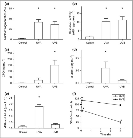
Oxidative damage induced by ultraviolet (UV) irradiation. Human melanocytes were exposed to UVA (60 J cm−2) or UVB irradiation (500 mJ cm−2). (a) Nuclear fragmentation quantified 24 h after irradiation and (b) caspase-3 activation detected 16 h after irradiation. AU, arbitrary units. Production of (c) cyclobutane pyrimidine dimers (CPDs) and (d) 8-hydroxydeoxyguanosine (8-OHdG) directly after irradiation. (e) Levels of the lipid peroxidation products malondialdehyde (MDA) and 4-hydroxyalkenals (4-HA) determined directly after irradiation. (f) Concentration of reduced glutathione (GSH) calculated as nmol mg−1 protein and expressed as the percentage of unirradiated cells from the same donor (set as 100%). Results are presented as the mean ± SD (n = 4). *P ≤ 0·05 vs. control.