
Figure 4.
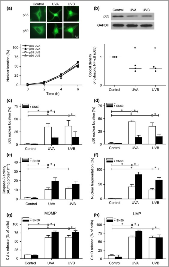
Nuclear factor (NF)-κB signalling in ultraviolet (UV)-irradiated melanocytes. Melanocytes were exposed to UVA (60 J cm−2) or UVB (500 mJ cm−2). (a) Immunostaining to determine the localization of the p50 and p65 subunits of the NF-κB dimer after irradiation. Images are selected to display the characteristic appearance of cytosolic and nuclear location, respectively. (b) One representative Western blot out of four and the corresponding optical density, showing the levels of the p65 subunit in digitonin-extracted cytosolic fractions. Glyceraldehyde-3-phosphate dehydrogenase (GAPDH) was used as an internal control. Nuclear localization of the (c) p65 and (d) p50 subunits in melanocytes pretreated with an inhibitor of the p50 subunit of the NF-κB dimer (SN50, 20 μmol L−1; black bars) 3 h prior to irradiation. (e) Caspase-3 activation was analysed based on the cleavage of the substrate Ac-DEVD-AMC, 16 h postirradiation [expressed as arbitary units (AU) per mg protein and hour], and (f) apoptosis was quantified via microscopic analysis of nuclear morphology in 4',6-diamidino-2-phenylindole (DAPI)-stained cells after 24 h. UV-induced (g) mitochondrial outer membrane permeabilization (MOMP), as detected by cytochrome c release, and (h) lysosomal membrane permeabilization (LMP), as detected by the release of cathepsin D (cat D). The results are presented as the mean ± SD (n = 4). *P ≤ 0·05 vs. control.