
Figure 6.
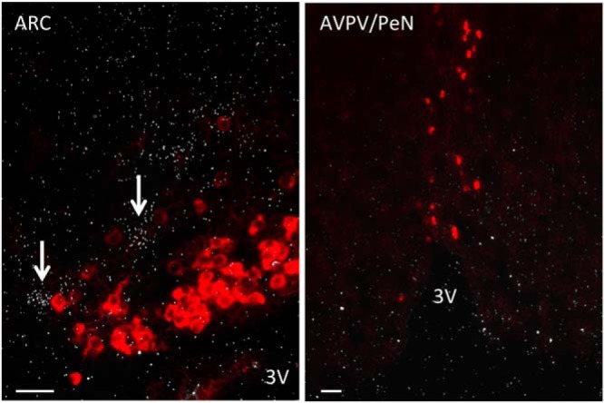
Representative microphotograph of a double label ISH depicting absence of colocalization between Kiss1-expressing neurons (red cells) and Tac1-expressing neurons (silver grains, indicated by white arrows) in the ARC (left panel) and AVPV/PeN (right panel) areas of adult OVX and OVX+E2 mice, respectively. 3V, third ventricle.