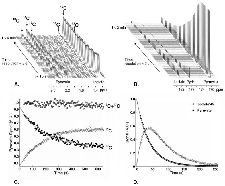
Figure 1.
(A) Real-time 1H-NMR and (B) hyperpolarized 13C-NMR assays of pyruvate-lactate exchange in colorectal SW1222 cancer cells and the corresponding time evolution of the spectral integrals (C, D). (C) Integrals of the 1H(12C) peak, open symbols, and 1H(13C) pyruvate peaks, closed symbols, from the 1H assay. (D) Integrals of the hyperpolarized [1-13C]pyruvate peak, closed symbols, and [1-13C]lactate peaks, open symbols, from the 13C assay. The solid lines correspond to the fits from the kinetic models described in the text.

