
Figure 4.
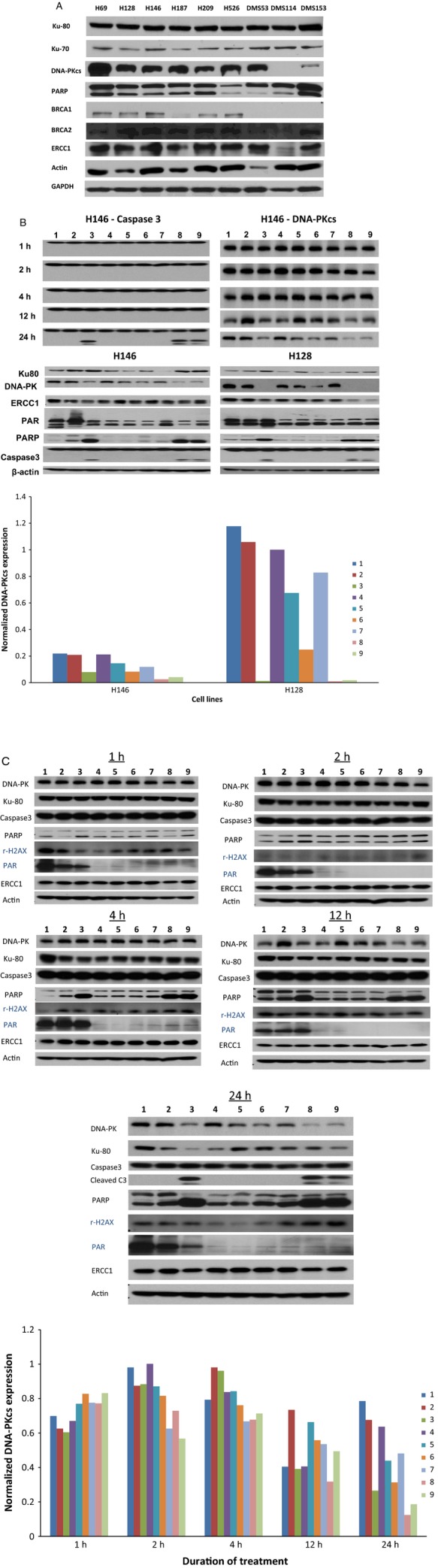
(A) Variable expression of BRCA1, ERCC1, and DNA-PKcs across the panel of SCLC cell lines. DNA-PKcs expression was noticeably high in cell lines (H69, H128 and DMS53) that were less sensitive to cisplatin or PARP inhibitor potentiation of cisplatin activity. (B) Top panel: Reduced expression of DNA-PKcs observed in H146 cell line coincided with onset of apoptosis as indicated by caspase 3 cleavage. Bottom Panel: DNA-PKcs expression was modulated in H146 and H128 cell lines but only at high concentrations of cisplatin and veliparib required for cytotoxicity (see bar graphs showing normalized DNA-PKcs expression relative to actin in H146 and H128 cell lines under different treatment conditions, 1–9, as measured by densitometry). There was no significant modulation of other representative DNA-repair enzymes. (C) Inhibition of PARP activity observed within 1 h of cell exposure to veliparib and persisted for the duration of the experiments 24 h later. Reduced DNA-PKcs expression observed at 24 h coincident with onset of apoptosis as indicated by cleaved caspase 3 and PARP (bar graphs showing normalized DNA-PKcs expression relative to actin in H146 cell lines as measured by densitometry under different treatment conditions, 1–9 and at different time points). Lanes: 1, Control; 2, cisplatin (2.5 μmol/L); 3, cisplatin (50 μmol/L); 4, veliparib (5 μmol/L); 5, veliparib (50 μmol/L); 6, cisplatin (2.5 μmol/L) + veliparib (5 μmol/L); 7, cisplatin (2.5 μmol/L) + veliparib (50 μmol/L); 8, cisplatin (50 μmol/L) + veliparib (5 μmol/L); 9, cisplatin (50 μmol/L) + veliparib (50 μmol/L).