
Figure 4.
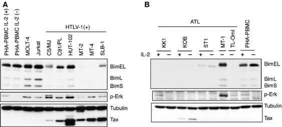
Expression of Bim in HTLV-1-infected T-cell lines. (A) Cell lysates were prepared from six HTLV-1-infected T-cell lines, two HTLV-1-uninfected T-cell lines and PHA-stimulated PBMC. The levels of Bim, p-Erk, Tax, and α-tubulin proteins were determined by western blot analysis using the corresponding antibodies. (B) Cell lysates were prepared from ATL-derived T-cell lines or PHA-PBMC cultured with or without IL-2 for 18 h. The levels of Bim, Tax, p-Erk, and α-tubulin proteins were determined by western blot analysis using the corresponding antibodies.