
Figure 5.
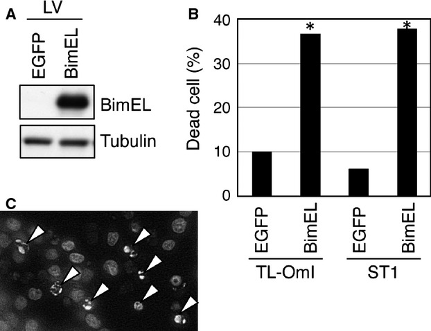
Re-expression of Bim in ATL cell lines induces apoptosis. (A, B, C) TL-OmI and ST1 cells were infected with lentivirus (LV) expressing Bim or control EGFP. Cell lysates were prepared from ST1 cells 24 h after infection, and the level of Bim protein was analyzed by western blotting (A). Cell viability 48 h after infection was evaluated by the trypan blue dye exclusion method (*P < 0.0001 by the Z-test) (B). Bim-transduced ST1 cells were evaluated for apoptosis by nuclear staining with Hoechst 33342. Arrows indicate the condensed nuclei of apoptotic cells (C). Data are representative of two independent experiments.