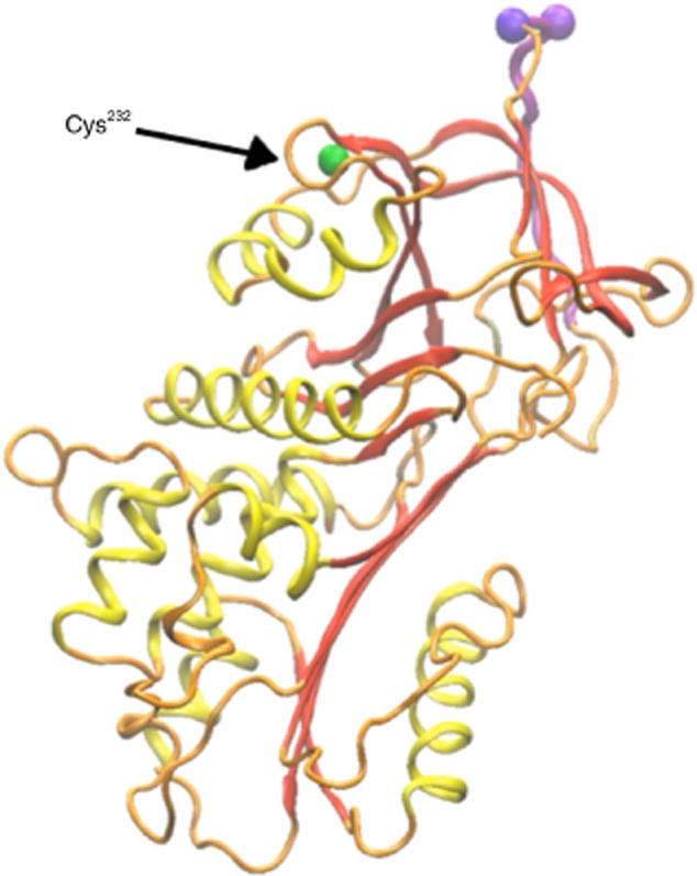
Figure 3.

In-silico depiction of the single surface cysteine residue within the sequence of α1-anti-trypsin (AAT). Orange = wire-diagram of the protein-sequence with secondary structures highlighted in yellow and red, and the protease-binding domain in purple. Non-exposed amino acids that are positioned under the surface of the molecule are represented by white beads. Green = cysteine at position 232.
