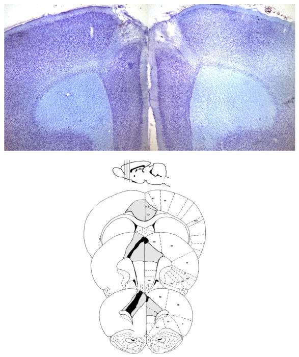
Figure 3.
ACC Histology. A) A representative image of the damage to the ACC at 2.7 mm anterior from bregma. The coronal section is 40 μm thick and stained with thionin. Damage was centered on rostral portions of the ACC and restricted to ACC. B) A diagram of the smallest (black) and largest lesions (gray) shows the regional selectivity of damage produced in the current study. All subjects included in the results sustained bilateral damage to the ACC.

