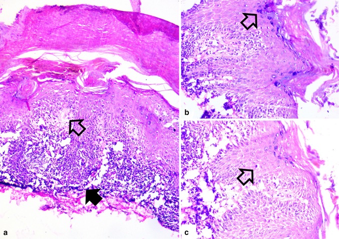
Fig. 2.
a Photomicrograph showing hyperkeratosis, basal vacuolar degeneration (open arrow), edema and prominent band like lymphocytic infiltrate (solid arrow) [100X] b higher magnification of wedge shaped hypergranulosis (arrow) with basal vacuolar degeneration [200X] and c occasional colloid bodies representing degenerating necrotic keratinocytes (arrow) [200X]

