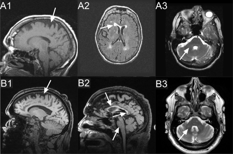
Figure 2.
Tesla MRI (1.5): T1 (A1), T2 FLAIR (A2), T2 (A3). 3 Tesla MRI: MPRAGE (B1), T2-FLAIR (B2), T2-TSE (B3); shown by arrows: Case 1: Moderate cerebral (A1) and mild cerebellar (not shown) volume loss, periventricular white matter lesions affecting anterior and posterior horns, bilaterally (A2). No white matter changes in the middle cerebellar peduncles (A3). Case 2: Moderate cerebral (B1), mild increased white matter changes in the middle cerebellar peduncles (B3) and pons (B2), moderately thin truncus of the corpus callosum (B2), with severe increased T2 signal intensity in both the truncus and the splenium of the corpus callosum (B2).

