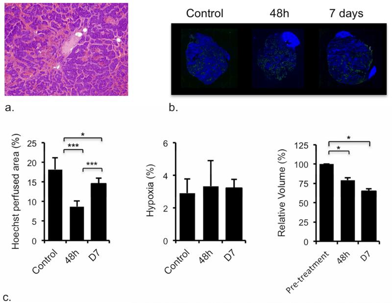
Figure 5.
Pathological response of TH-MYCN model of neuroblastoma to cediranib. a) Microscopic image (×40) from a hematoxylin and eosin stained section prepared from a tumor of a TH-MYCN mouse, 48h after treatment with cediranib, showing increased vascular thrombosis (c.f. control tumor in Figure 2c). The frequency of vascular thrombosis was significantly greater in sections from the cediranib tumors compared with control (p=0.03, χ2-test, n=4 treated, n=7 control). b) Composite fluorescence images of uptake of the perfusion marker Hoechst 33342 (blue), and pimonidazole adduct formation (green) into the tumor and kidneys of TH-MYCN mice, demonstrating reduced vascular perfusion (expressed as Hoechst perfused area) in tumors of TH-MYCN mice, 48h and 7 days after treatment with a daily dose of 6mg/kg cediranib, compared with tumors of control TH-MYCN mice (*p<0.05, **p<0.01, ***p<0.005, Student’s 2-tailed unpaired t-test). The degree of tumor hypoxia was negligible, and no differences in hypoxia between treated and control tumors were detected. Tumor volumes decreased over 48h in tumor-bearing TH-MYCN mice, an effect sustained at 7 days following daily treatment with 6mg/kg cediranib. (*p<0.05, **p<0.01, ***p<0.005, Student’s 2-tailed paired t-test).

