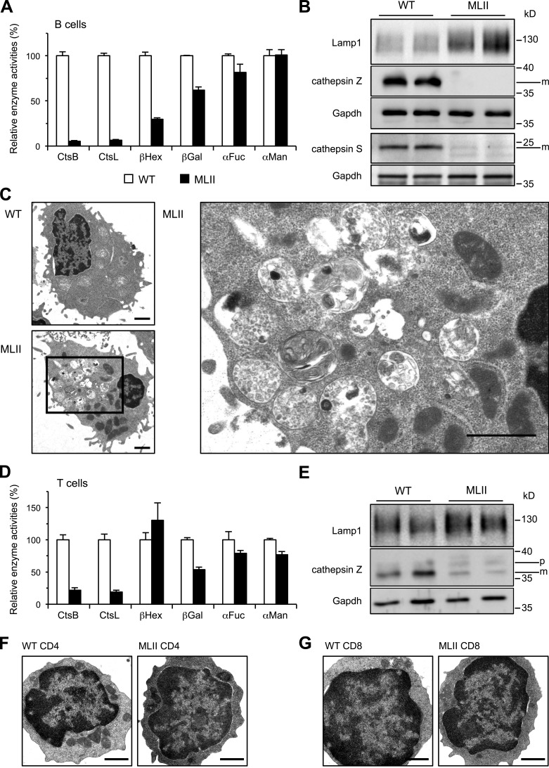
Figure 1.
Loss of lysosomal proteases and accumulation of storage material in B cells from MLII mice. (A) The activities of CtsB, CtsL, βHex, βGal, αFuc, and αMan in extracts of B cell blasts. Specific activities in B cells of WT mice were set to 100%. Mean and SEM (n = 3–6). (B) Western blot from B cell extracts. (C) Electron micrographs of splenic B cell blasts revealed accumulation of lysosomal storage material in MLII cells. Higher magnification of indicated area from a representative MLII B cell is shown. Bars, 1 µm. (D) Relative activities of CtsB, CtsL, βHex, βGal, αFuc, and αMan in extracts of T cell blasts (percentage of WT controls). Mean and SEM (n = 3–6). (E) Western blot from T cell extracts. m, mature form; p, precursor form. (F and G) Electron micrographs of purified splenic MLII CD4 T cells (F) and CD8 T cells (G) revealed normal appearance. Bars, 1 µm.

