Figure 2.
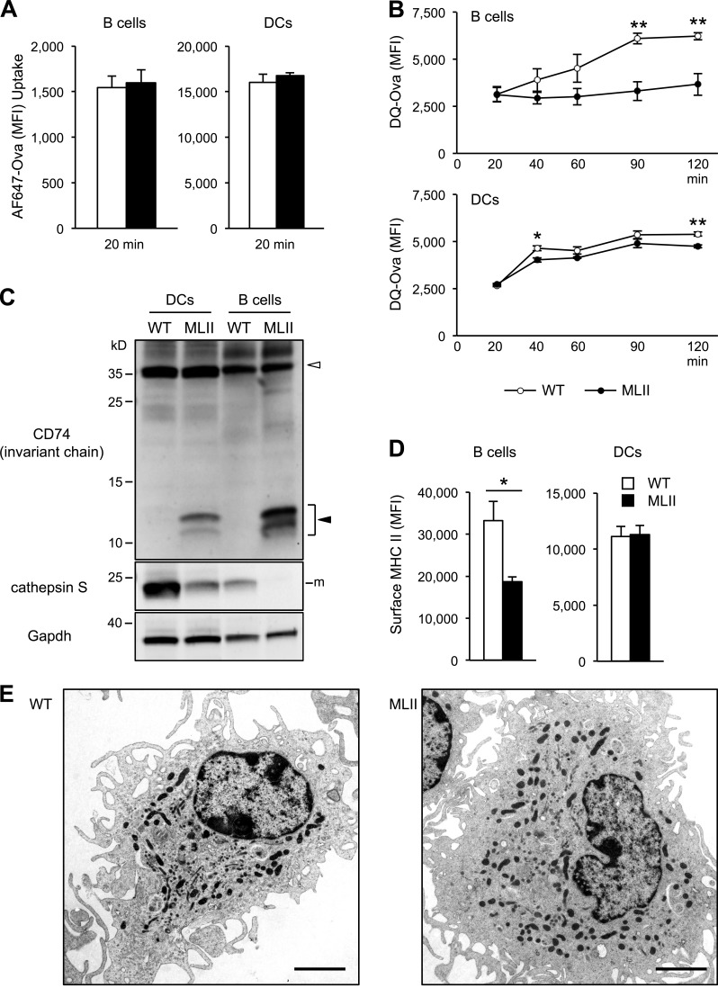
Cathepsin deficiencies impair antigen and CD74 degradation in antigen-presenting cells from MLII mice. (A) Uptake of Alexa Fluor 647 (AF647)–Ova in WT and MLII B cells and DCs for 20 min. (B) Fragmentation of DQ-Ova antigen in B cells and DCs. After uptake the proteolytic release of DQ fluorescence was quantified over time. (C) Representative CD74 and CtsS Western blot from WT and MLII B cells and DCs. Positions of full-length CD74 (open arrowhead) and of accumulating CD74 degradation intermediates (closed arrowhead) are indicated. m, mature form. (D) Total surface MHC II level on B cells and DCs analyzed by flow cytometry. Mean and SEM (n = 3). *, P < 0.05; **, P < 0.01. (E) Electron micrographs of MLII DCs revealed normal appearance. Bars, 2.5 µm.

