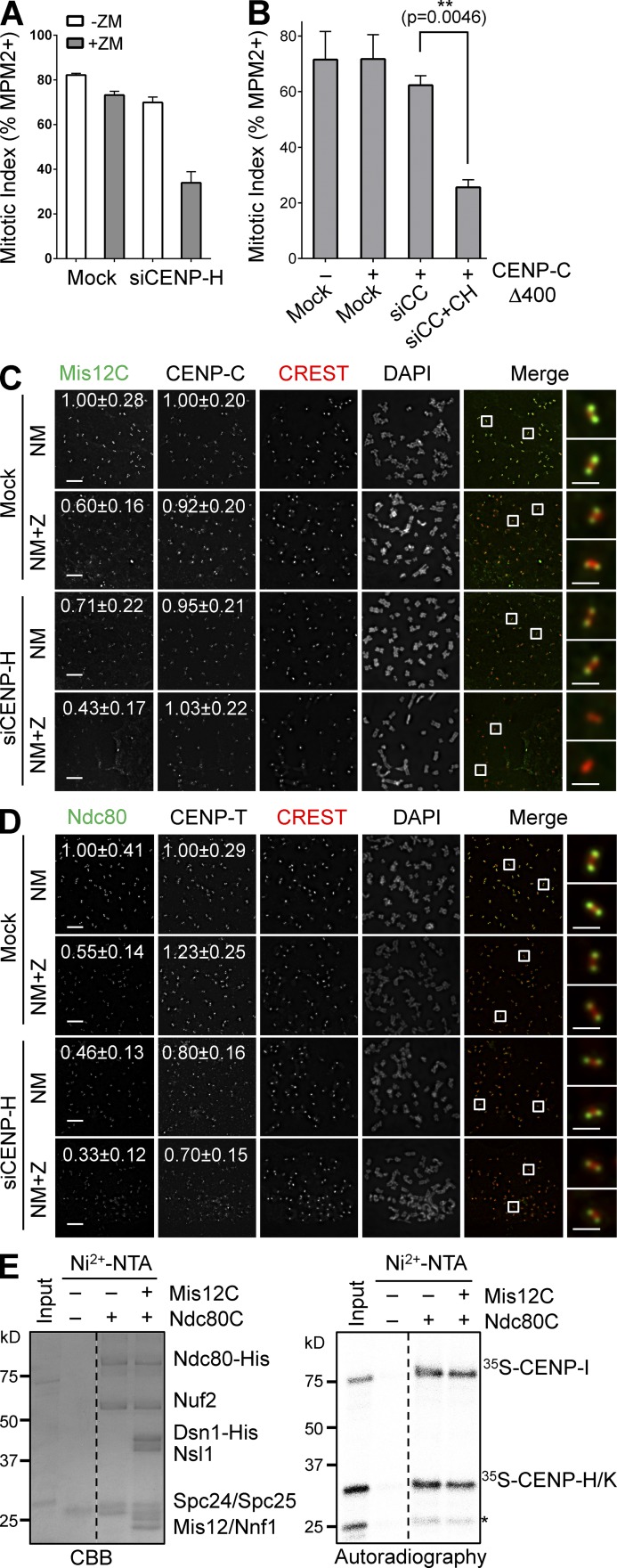
Figure 6.
CENP-T promotes KMN kinetochore targeting through CENP-H-I-K. (A) HeLa Tet-On cells were mock transfected or transfected with siCENP-H, treated with thymidine for 14 h, released into nocodazole-containing medium for 12 h, and treated with or without ZM for 2 h. Their mitotic index was determined by flow cytometry. Means and SDs (error bars) are shown (n = 3 independent experiments). (B) HeLa cells were transfected with the indicated plasmids and siRNAs (siCC, siCENP-C; siCC+CH, siCENP-C+siCENP-H), and treated with nocodazole. Their mitotic index (mean ± SD [error bars], n = 3) was quantified by flow cytometry. (C and D) Nocodazole-treated mitotic HeLa cells transfected with siCENP-H were further incubated with MG132 (NM) or with both MG132 and ZM (NM+Z), and stained with the indicated antibodies and DAPI. Boxed regions of merged images were magnified and shown in the rightmost column. The relative kinetochore intensities (mean ± SD, n = 400) in certain channels were quantified and shown. Bars, 5 µm (1 µm for magnified images). (E) Recombinant Ndc80C was preincubated with or without recombinant Mis12C, immobilized on beads, and incubated with 35S-labeled CENP-H-I-K. Bound proteins and input were separated by SDS-PAGE, stained with CBB (left), and analyzed with a phosphorimager (right). CENP-H and -K co-migrate. The asterisk indicates a CENP-K fragment. Broken lines indicate that intervening lanes have been spliced out.

