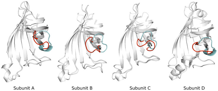
Figure 16. The final simulated aMD structure for tetramer streptavidin is superimposed onto the crystal structure.

Four chains are shown as Subunit A, Subunit B, Subunit C and Subunit D, respectively. Cyan color stands for the conformation of the loop in the open crystal structure, red color for the simulated structure.
