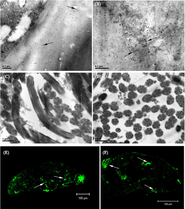
Figure 5. Immunolabelling micrographs showing the distribution of RSV RNP particles in SBPH germ cells.
A–D are transmission electron micrographs, and E and F are confocal laser micrographs. A: RSV RNP particles on the eggshell; B: RSV RNPs in the interior of ovum; C and D: the sperms of viruliferous SBPH without virus infection; the transverse section of sperms was approximately circular, and the longitudinal section was cylindrical shape; E and F: RSV RNPs in the interior of eggs after spawning. Virus RNPs were indicated by arrows. Scale bars: 0.2 μm (A, B), 1 μm (C, D) and 100 μm (E, F).

