Figure 1.
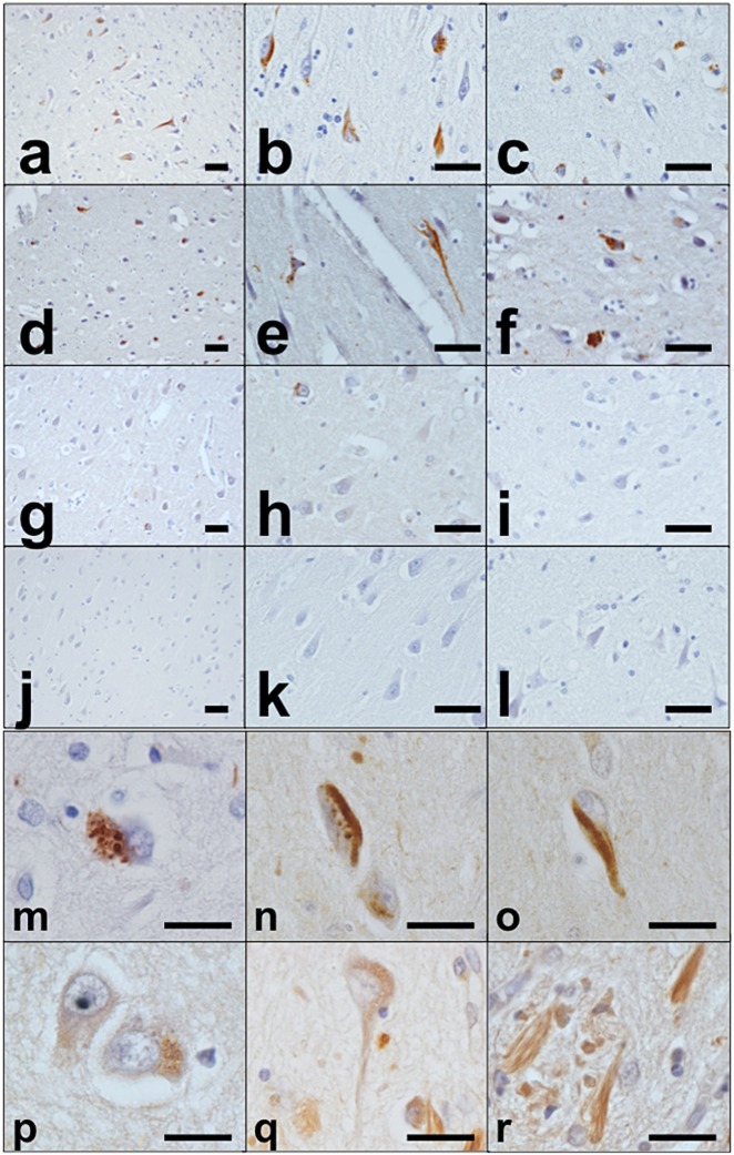
Immunohistochemistry for PtdIns(4,5)P2. Immunohistochemistry in AD, MyD, ALS and control cases. AD case (case 1): low-power fields in hippocampus (a,b) and neocortex (c). MyD case (case 6): low-power fields in hippocampus (d,e) and neocortex (f). ALS case (case 17): low-power fields in hippocampus (g,h) and neocortex (i). Control case (case 26): low-power fields in hippocampus (j,k) and neocortex (l). AD case (case 5): some of the detected structures are (m) GVD bodies, (n) ‘GVD bodies + NFTs’, and (o) NFTs. The specificity of immunoreactivity for PtdIns(4,5)P2 was verified using PLCδ1PH-GST on sections, which showed essentially the same staining pattern as the anti-PtdIns(4,5)P2 antibody (p–r). Scale bars: (a–l) 50 μm, (m–r) 20 μm.

