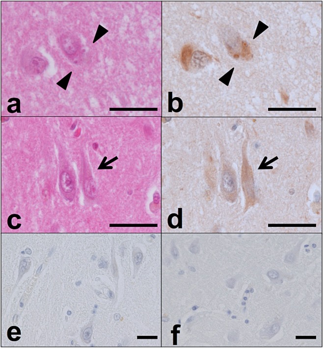
Figure 2.
PtdIns(4,5)P2-positive structures corresponding to GVD bodies and NFTs. In the hippocampus of an AD case, the cellular localization of PtdIns(4,5)P2 was compared among haematoxylin and eosin (H&E)–stained sections (a,c), sections subjected to immunohistochemical analysis with 2C11 [b,d; some of the structures are GVD bodies (arrowhead) and some are NFTs (arrow)], sections incubated without primary antibody (e), and sections incubated with 10 mM neomycin sulphate/PBS prior to immunohistochemical staining with 2C11 (f). Scale bars: 20 μm.

