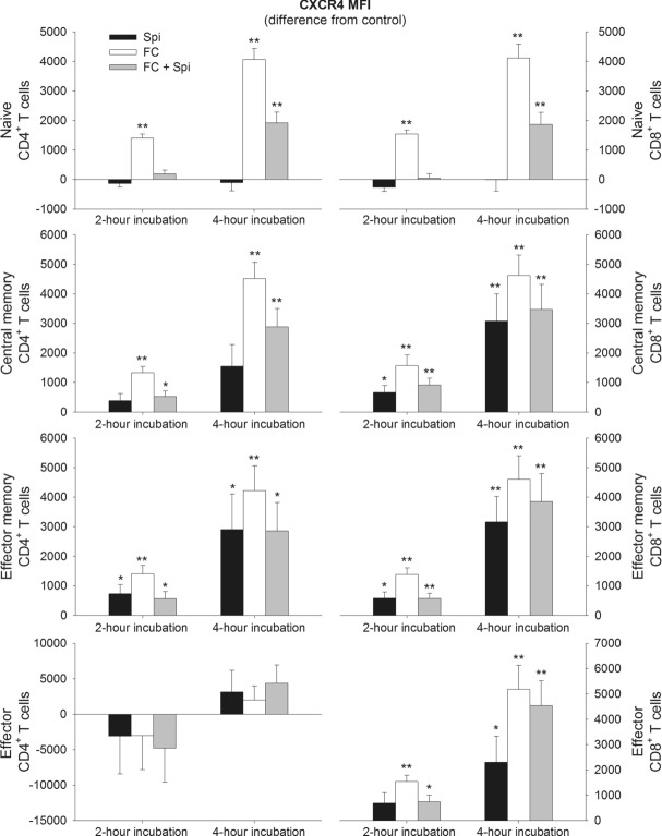
Figure 5.
Changes in CXCR4 expression on CD4+ and CD8+ T-cell subsets after incubation with spironolactone and fludrocortisone. Blood was sampled during sleep at 03:30 h and was then incubated for 2 or 4 h either with spironolactone (Spi), fludrocortisone (FC), fludrocortisone plus spironolactone (FC + Spi) or PBS as control. Expression of CXCR4 was assessed by flow cytometry on naïve (CD45RA+CD62L+), central memory (CD45RA−CD62L+), effector memory (CD45RA−CD62L−) and effector (CD45RA+CD62L−) CD4+ (left) and CD8+ (right) T cells and is shown as mean ± SEM of median fluorescence intensity (MFI) of 13 healthy male subjects. Data are expressed as difference from the PBS control. *p < 0.05, **p < 0.01, for pairwise comparisons between effects of the active agent(s) and PBS (paired t-tests).

