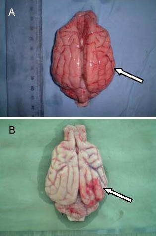
Figure 1.

Gross specimens in the treatment group and the control group at 24 hours after cerebral infarction in Beagle dogs.
In the control group (B), the number of thickened blood vessels increased at the peripheral infarct site. In the treatment group (A), hyperplasia was reduced. Arrows indicate blood vessels at the infarct site.
